

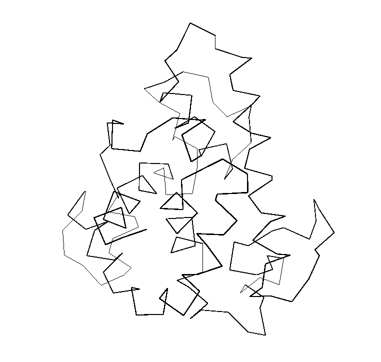
 Figure 1. (left) XmMol representation of 153l,
(right) XmMol representation of 153l using superimposed Protein Blocks.
Figure 1. (left) XmMol representation of 153l,
(right) XmMol representation of 153l using superimposed Protein Blocks.
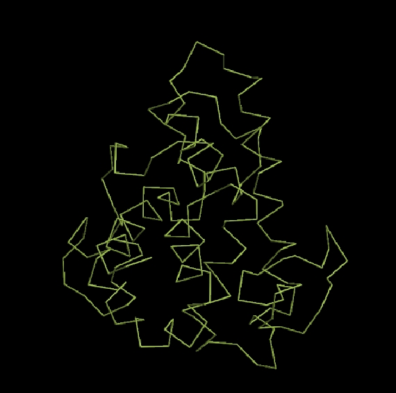
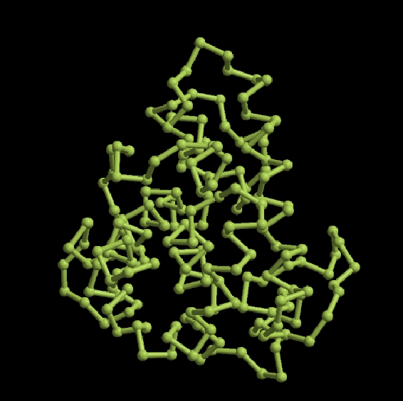 Figure 2. (left) raster 3D representation of 153l,
(right) raster 3D representation of 153l.
Figure 2. (left) raster 3D representation of 153l,
(right) raster 3D representation of 153l.
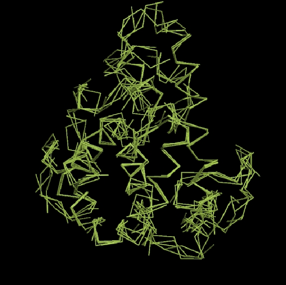
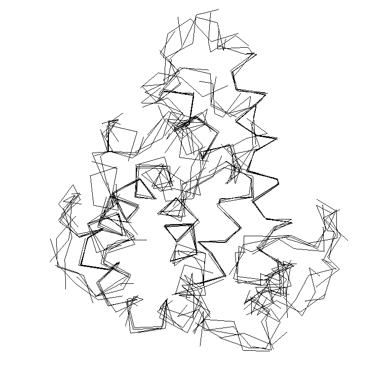
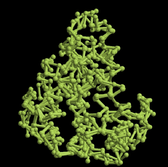 Figure 3. (left) raster 3D representation of 153l using superimposed Protein Blocks,
(right) raster 3D representation of 153l using superimposed Protein Blocks.
Figure 3. (left) raster 3D representation of 153l using superimposed Protein Blocks,
(right) raster 3D representation of 153l using superimposed Protein Blocks.
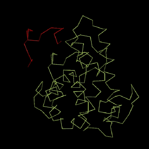
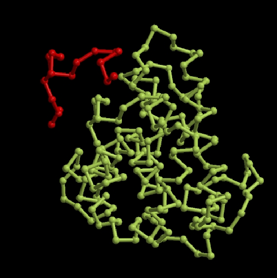 Figure 4. (left and right) 3D structure of protein 153l in green and in red, the N-ter of 153l,
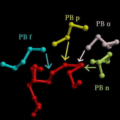
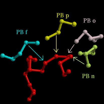 Figure 4. (left and right) in red, the N-ter of 153l and the Protein Blocks associated with the first residues.
Top
back
Last modif : 25 April 2004