INSERM EBGM U436

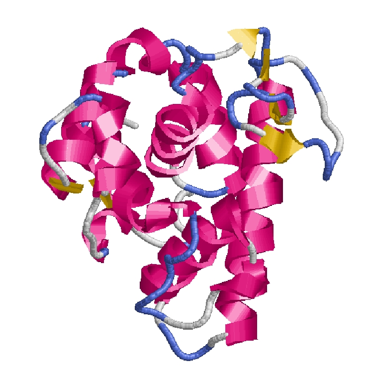
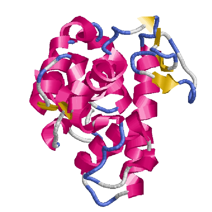
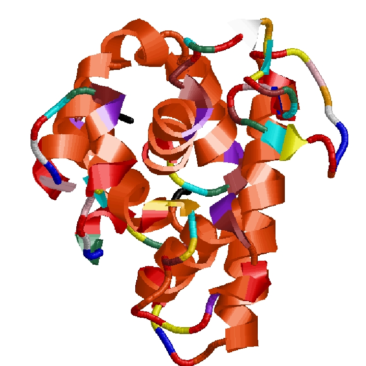
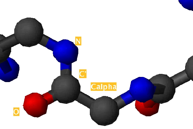
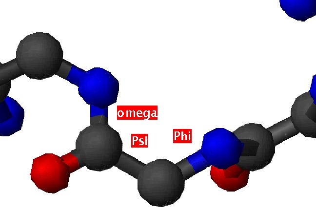
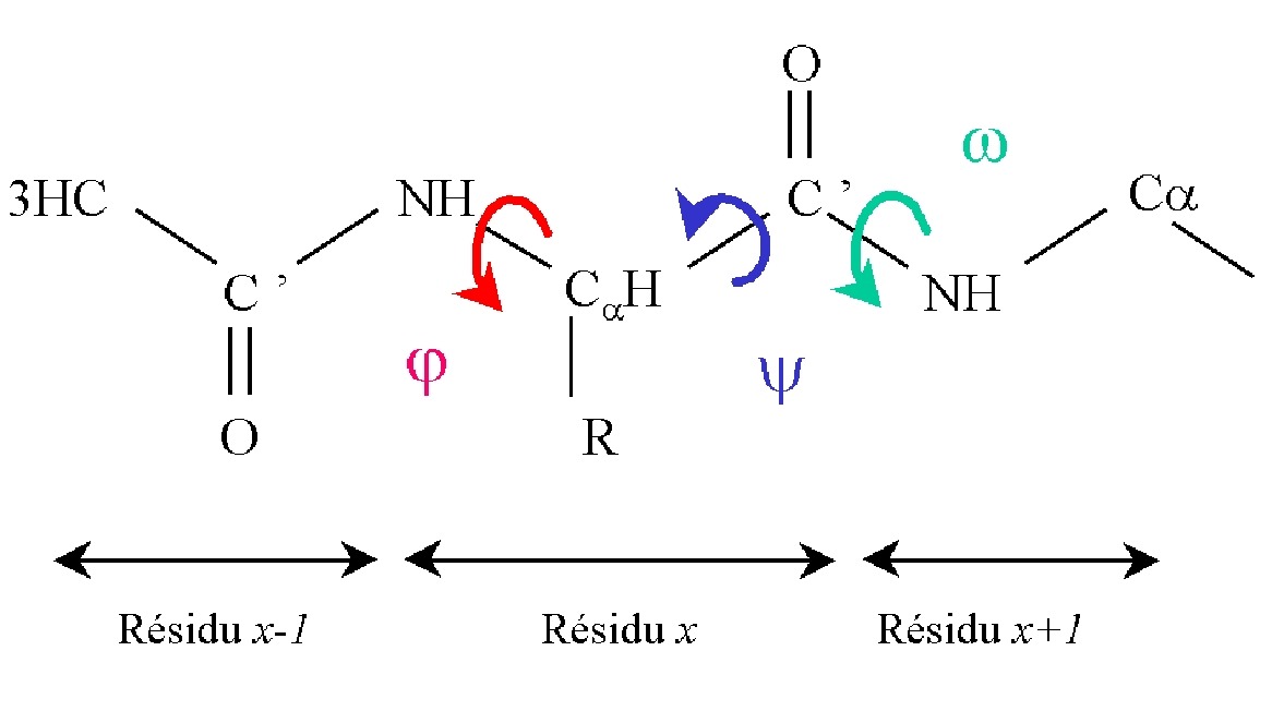
 How is a 3D protein structure coded into Protein Blocks ?

anim 09


Proteins are composed of an amino acid succession and could be easily seen with different representations (see Figure 1 ).
protein protein

1.Protein figures.

Two protein representations of protein 153L (left) : ribbons derived from secondary structures and (right) : cpk models (both molmol software).

Top


2. PDB file.

Proteins structure are mainly described in PDB format (and mCIF). The PDB format is composed of different xxxx. The most interesting is the "ATOM" xxxx which contains the type of every atom resolved with their X, Y and Z positions.


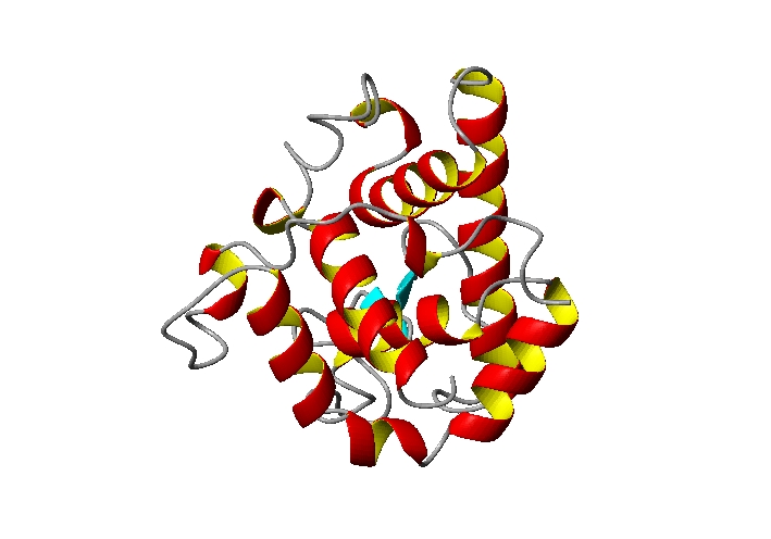
	HEADER    HYDROLASE(O-GLYCOSYL)                   05-MAY-94   153L      153L   2
	ATOM      1  N   ARG     1       6.350  34.124  50.750  1.00 41.90      153L 116
	ATOM      2  CA  ARG     1       6.324  32.707  50.379  1.00 54.68      153L 117
	ATOM      3  C   ARG     1       6.334  32.484  48.874  1.00 14.63      153L 118
	ATOM      4  O   ARG     1       7.356  32.060  48.316  1.00 23.75      153L 119
	ATOM      5  CB  ARG     1       5.009  32.300  50.934  1.00 40.20      153L 120
	ATOM      6  CG  ARG     1       4.526  33.584  51.604  1.00 56.81      153L 121
	ATOM      7  CD  ARG     1       3.012  33.793  51.724  1.00 64.62      153L 122
	ATOM      8  NE  ARG     1       2.515  34.238  50.431  1.00 63.14      153L 123
	ATOM      9  CZ  ARG     1       2.352  33.394  49.423  1.00 37.38      153L 124
	ATOM     10  NH1 ARG     1       2.588  32.086  49.557  1.00 56.33      153L 125
	ATOM     11  NH2 ARG     1       1.895  33.858  48.262  1.00 59.78      153L 126
	ATOM     12  N   THR     2       5.206  32.767  48.261  1.00 15.22      153L 127
	ATOM     13  CA  THR     2       5.197  32.618  46.826  1.00 15.40      153L 128
	ATOM     14  C   THR     2       4.781  33.870  46.108  1.00 23.28      153L 129
	ATOM     15  O   THR     2       4.716  33.930  44.845  1.00 20.96      153L 130
	ATOM     16  CB  THR     2       4.452  31.426  46.229  1.00 12.40      153L 131
	ATOM     17  OG1 THR     2       3.089  31.502  46.538  1.00 20.80      153L 132
	ATOM     18  CG2 THR     2       5.066  30.133  46.701  1.00 26.07      153L 133
	ATOM     19  N   ASP     3       4.497  34.900  46.908  1.00 31.19      153L 134
	ATOM     20  CA  ASP     3       4.020  36.132  46.259  1.00 35.11      153L 135
	ATOM     21  C   ASP     3       4.987  37.273  45.992  1.00 19.94      153L 136
	ATOM     22  O   ASP     3       4.530  38.398  45.795  1.00 29.83      153L 137
	ATOM     23  CB  ASP     3       2.951  36.717  47.185  1.00 28.17      153L 138
	ATOM     24  CG  ASP     3       3.632  36.891  48.524  1.00 26.63      153L 139
	ATOM     25  OD1 ASP     3       4.821  36.990  48.715  1.00 43.06      153L 140
	ATOM     26  OD2 ASP     3       2.844  36.730  49.510  1.00 54.69      153L 141
	ATOM     27  N   CYS     4       6.285  37.076  46.089  1.00 17.27      153L 142
	ATOM     28  CA  CYS     4       7.131  38.210  45.919  1.00 16.83      153L 143
	ATOM     29  C   CYS     4       7.073  38.939  44.605  1.00 15.34      153L 144
	ATOM     30  O   CYS     4       7.459  40.095  44.530  1.00 22.22      153L 145
	ATOM     31  CB  CYS     4       8.597  37.815  46.045  1.00 14.54      153L 146
	ATOM     32  SG  CYS     4       8.982  37.124  47.655  1.00 23.95      153L 147
	ATOM     33  N   TYR     5       6.691  38.264  43.538  1.00 11.91      153L 148
	ATOM     34  CA  TYR     5       6.719  38.935  42.270  1.00 13.01      153L 149
	ATOM     35  C   TYR     5       5.366  39.179  41.653  1.00 13.67      153L 150
	ATOM     36  O   TYR     5       5.254  39.476  40.486  1.00 19.57      153L 151
	ATOM     37  CB  TYR     5       7.599  38.124  41.282  1.00 21.26      153L 152
	ATOM     38  CG  TYR     5       9.055  37.997  41.772  1.00 17.97      153L 153
	ATOM     39  CD1 TYR     5       9.974  39.045  41.627  1.00 28.24      153L 154
	ATOM     40  CD2 TYR     5       9.485  36.843  42.432  1.00 16.83      153L 155
	ATOM     41  CE1 TYR     5      11.281  38.964  42.115  1.00 29.20      153L 156
	ATOM     42  CE2 TYR     5      10.780  36.732  42.944  1.00 19.39      153L 157
	ATOM     43  CZ  TYR     5      11.672  37.794  42.763  1.00 27.35      153L 158
	ATOM     44  OH  TYR     5      12.956  37.704  43.225  1.00 26.92      153L 159
	ATOM     45  N   GLY     6       4.320  39.020  42.427  1.00 19.79      153L 160
	ATOM     46  CA  GLY     6       2.986  39.221  41.892  1.00 15.13      153L 161
	ATOM     47  C   GLY     6       2.071  38.002  41.992  1.00 17.68      153L 162
	ATOM     48  O   GLY     6       2.522  36.960  42.438  1.00 21.96      153L 163
	ATOM     49  N   ASN     7       0.819  38.158  41.541  1.00 18.97      153L 164
	ATOM     50  CA  ASN     7      -0.269  37.184  41.565  1.00 20.89      153L 165
	ATOM     51  C   ASN     7      -0.922  37.069  40.232  1.00 13.79      153L 166
	ATOM     52  O   ASN     7      -1.584  37.990  39.760  1.00 14.39      153L 167
	ATOM     53  CB  ASN     7      -1.372  37.625  42.536  1.00 19.21      153L 168
	ATOM     54  CG  ASN     7      -2.418  36.535  42.784  1.00 42.06      153L 169
	ATOM     55  OD1 ASN     7      -2.908  35.750  41.931  1.00 29.02      153L 170
	ATOM     56  ND2 ASN     7      -2.761  36.491  44.049  1.00 54.80      153L 171
	END                                                                     153L1736
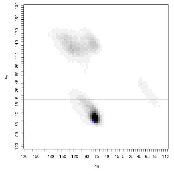
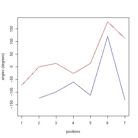
Figure 2 is an example of the PDB file format with the N-t of protein 153l.

Top

3. Protein backbone.

So a protein is defined by both the backbone (succession of N, Calpha, C and 0) common to every type of residues and the side-chains of every residues. The side-chains are essential for the interactions of the proteins, the backbone reflects the curve of the protein. In our study, we have focussed on the backbone.

protein

Figure 3. N-t of protein 153l with both backbone and side-chains.

protein

Figure 4. N-t of protein 153l with only the protein backbone.

Top

4. Protein backbone: atoms and angles.

The backbone is composed of 4 atoms describing the peptide plan : two consecutive carbones names Calpha and C', an azote (N) and an oxygen (O). The oxygen does participate directly in the peptide bond .

protein

Figure 5. Zoom on the protein backbone.
protein        protein

Figure 6. (left) atoms defining the protein backbone and (right) the different angles.
diehdral angles : phi , psi et omega, describe the protein backbone. By taking into account for the angle phi x (phi in position x in the sequence), atoms C'x-1, Nx, C alpha x and C'x, for psi x, atoms Nx, C alpha x and C'x et Nx+1 and for omega, C alpha x, C'x, Nx+1 and C alpha x+1.

diehedral angles        Ramachandran

Figure 7. (left) definition of Phi, Psi and omega angles and (right) classical distribution of Phi-Psi angles using Ramachandran dotplot.
Top

5. Coding of the 3D structures: use of Phi - Psi angles.

So to describe the 3D protein structure, we have use the succession of Phi - Psi angles. Here, it is an exmple of the traduction of the 3D (X,Y,Z) into Phi-Psi vectors with the example of protein 153l.

Position Phi Psi
1 --- -73.17
2 -124.40 0.21
3 -98.44 13.49
4 -60.37 -25.89
5 -112.84 13.68
6 120.99 178.29
7 -130.63 112.67
Figure 8. Traduction of protein 153l N-t into Phi / Psi (cf. Figure 2).

protein        protein

Figure 9. Representation of Phi - Psi vectors for the (left) 153l N-t and complete 153l (right), with phi in blue and psi in red.
Top

6. Coding in terms of Protein Blocks : example.

So, the angular vectors are translated into Protein Blocks. Each angular vectors is compared to the 16 Protein Blocks angular vectors (defined in the learning step) and is assigned to the one which it is the close. The metric is simple, it is called Root Mean Square deviation on angular values (noted RMSda). A vector is assigned to the Protein Blocks which share the minimal RMSda.

raster 3D 153l and Nter raster 3D 153l Nter and PBs

Figure 10. (left) 3D structure of protein 153l in green and in red, the N-ter of 153l,

(right) in red, the N-ter of 153l and the Protein Blocks associated with the first residues (click here for other representations).

Top

7. Coding in terms of Protein Blocks : results.

Here is presented the coding of 3D structure of protein 153l, both in terms of Protein Blocks and classical secondary structures. The Figures 11 and 12 show the interest of using the Protein Blocks instead of secondary structures to describe all the protein 3D structures.

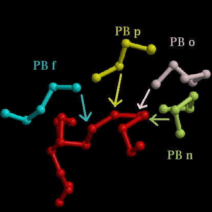
	    1
	(A) RTDCYGNVNRIDTTGASCKTAKPEGLSYCGVSASKKIAE
	(B) ZZmnopfklpccebjafklmmmnopabecjklmmmmmmm
	(C) cccccccccccccccccaaaaaaaccccccaaaaaaaaa
	(D) CCTTTTCGGGCCCCCBCHHHHGGGCCCCCBHHHHHHHHH

	    41
	(A) DLQAMDRYKTIIKKVGEKLCVEPAVIAGIISRESHAGKV
	(B) mmmmmmmmmmmmmmmmmnopafklmmmmmmmmnooolap
	(C) acaaaaaaaaaaaaaaaaacccaaaaaaaaaaacccccc
	(D) HHHHHHHHHHHHHHHHHHHCCCHHHHHHHHHHHHGGGTT

	    81
	(A) KNGWGDRGNGFGLMQVDKRSHKPQGTWNGEVHITQGTTI
	(B) ehiafkopagcjkopafklmccehjfklmklmmmmmmmm
	(C) cccccccccccccccccccccccccccccaaaaaaaaaa
	(D) BTTBTTTTCEETTTTEETTTTCCCCTTTTHHHHHHHHHH

	    121
	(A) INFIKTIQKKFPSWTKDQQLKGGISAYNAGAGNVRSYAR
	(B) mmmmmmmmmmmmbcfklmmmmmmmmmmnomklmnbfklm
	(C) aaaaaaaaaacccccaaaaaaaaaaaaaccccccccccc
	(D) HHHHHHHHHHTTTTCHHHHHHHHHHHHHHCGGGCCTTTT

	    161
	(A) DIGTTHDDYANDVVARAQYYKQHGY
	(B) goiahilmmmmmmmmmmmmmmnoZZ
	(C) ccccccccaaaaaaaaaaaaaaacc
	(D) TTTTTTTTHHHHHHHHHHHHHHCCC
Figure 10. (A) amino acid sequence, (B) Protein Blocks traduction, (C) secondary structures determined by P-SEA software and (D) by STRIDE software.
protein in terms of Protein Blocks        protein in terms of Protein Blocks
Figure 11. Rasmol visualization of protein 153l with secondary structures.
protein in terms of Protein Blocks        protein in terms of Protein Blocks
Figure 12. Rasmol visualization of protein 153l with Protein Blocks colors.
Top

7. Conclusion.

Hence from the 3D structures, the 3D structures are more precisely describded than with the classical secondary structure which let 50% of the structures unassigned (the coils). Other examples of coded proteins are given.

For more details on the Protein coding, please mail me at debrevern@urbb.jussieu.fr.

Top
back
Last modif : 25 April 2004